Catheter-based AI imaging is a groundbreaking advancement transforming cardiovascular diagnostics. The incorporation of miniature imaging devices into catheters coupled with AI algorithms offer clinicians unparalleled insights into coronary arteries, setting a new standard in detecting and managing CAD.
The Evolution of Miniaturized Catheter Technology
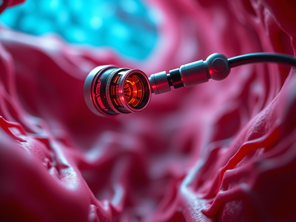
The Evolution of Miniaturized Catheter Technology is a pivotal development in the realm of cardiovascular diagnostics, radically transforming the approach to detecting and managing coronary artery disease (CAD). At the heart of this revolution lies the integration of cutting-edge advancements in hybrid imaging catheters, nano-optics, advanced biocompatible materials, and antimicrobial coatings. These innovations are propelling the creation of ultraflexible, micro-scale catheters that navigate the cardiovascular system with unprecedented precision and safety.
Hybrid imaging catheters stand as a cornerstone of this technological leap. By combining multiple imaging modalities, such as optical coherence tomography (OCT) and intravascular ultrasound (IVUS) within the same device, clinicians can now obtain a comprehensive view of the coronary arteries. This dual-modality approach enriches the diagnostic process, providing both the high-resolution detail of OCT and the deep tissue penetration of IVUS. The integration of nano-optics into these catheters further enhances their imaging capabilities, enabling the resolution of structures at the nanometer scale. This level of detail is critical in identifying early signs of plaque build-up and vessel wall abnormalities, factors that traditional imaging techniques might overlook.
Advancements in biocompatible materials are also critical to the evolution of miniaturized catheter technology. Modern catheters are constructed from materials that not only ensure the device’s longevity within the harsh biochemical environment of the body but also minimize the risk of adverse reactions. These materials are tailored to provide the perfect balance of flexibility and strength, ensuring that catheters can navigate through tight and tortuous vessels without causing damage to the vessel walls.
The threat of infection is a significant concern with any invasive procedure. To address this, the latest catheters are being coated with advanced antimicrobial agents that inhibit bacterial growth, thereby substantially reducing the risk of post-procedural infections. This innovation is particularly pertinent in the era of increasing antibiotic resistance, offering an added layer of protection to patients undergoing diagnostic catheterizations.
Robotic navigation and AI-assisted steering mechanisms represent another transformative development in catheter technology. These systems enable precise, semi-automated navigation through the complex architecture of the heart and blood vessels, significantly reducing the physical strain on the operator and enhancing the safety and accuracy of the procedure. For example, catheters equipped with magnetic tips can be steered with incredible precision using external magnetic fields, allowing them to reach areas of the cardiovascular system that were previously inaccessible.
This revolution in catheter technology is not occurring in isolation; it is part of a broader trend toward the integration of AI and robotic systems in cardiovascular diagnostics. The application of AI algorithms for real-time imaging data analysis and the adoption of robotic assistance for procedural navigation are streamlining diagnostics, refining treatment planning, and enhancing patient safety. These technologies are setting new standards for what is possible in the detection and management of coronary artery disease, marking a significant shift away from traditional methods and towards a future where cardiovascular conditions can be diagnosed and treated with unparalleled precision and personalization.
In conclusion, the advancements in miniaturized catheter technology, from hybrid imaging capabilities and nano-optics integration to the application of advanced materials and coatings, are revolutionizing the landscape of cardiovascular diagnostics. By providing clinicians with tools that offer superior imaging resolution, flexibility, and safety, this revolutionary technology is paving the way for more accurate, efficient, and personalized approaches to diagnosing and managing cardiovascular diseases.
AI-Assisted Imaging and Diagnostic Breakthroughs
AI-assisted imaging and diagnostic breakthroughs are dramatically reshaping the landscape of cardiovascular medicine, leveraging the precision of artificial intelligence to enhance imaging, diagnostics, and patient care. Building on the advancements in miniaturized catheter technology discussed in the previous chapter, AI integration takes cardiovascular diagnostics to new heights. This fusion of technology enables clinicians to detect, diagnose, and manage coronary artery disease (CAD) with unparalleled accuracy and efficiency.The integration of AI algorithms with intravascular imaging data marks a significant leap forward in the detection of subtle, previously invisible signs of early coronary artery disease. AI-enhanced imaging lenses within catheters can now provide real-time, high-resolution insights into the vascular system, allowing for the identification of early-stage abnormalities that might be missed by traditional imaging techniques. This capability is critical in planning and executing personalized treatment strategies, particularly during percutaneous coronary interventions (PCI), where immediate feedback on vessel morphology and physiology is invaluable.
One of the key benefits of AI integration in cardiovascular diagnostics is the streamlining of diagnostic workflows in catheterization labs. By analyzing complex imaging data in real-time, AI algorithms can quickly identify patterns indicative of CAD, significantly reducing the time doctors spend interpreting data. This efficiency not only accelerates the diagnostic process but also reduces the potential for human error, ensuring that treatment decisions are based on the most accurate and comprehensive information available.
Moreover, the advent of robotic and AI-assisted navigation represents a quantum leap in the safety and precision of cardiovascular procedures. Robotic systems equipped with AI-enhanced imaging capabilities can navigate the cardiovascular system with unprecedented precision, allowing for semi-automated imaging that reduces operator fatigue and minimizes the risk of procedural complications. This technological synergy ensures that patients receive the most accurate diagnoses and effective treatments with minimal invasiveness.
Beyond catheter-based imaging, AI is revolutionizing cardiovascular diagnostics across a broad spectrum of applications. For instance, AI-based ECG analysis has shown remarkable success in predicting cardiovascular risks and detecting conditions much earlier than traditional methods. This early detection is crucial for timely intervention and can significantly improve patient outcomes. Similarly, AI’s role in enhancing the diagnostic power of multimodal imaging, including CT scans and MRI, is becoming increasingly important. Cutting-edge technologies like Cleerly’s AI-driven quantitative CT (AI-QCT) and GE HealthCare’s Revolution Vibe CT system are setting new standards for accuracy in cardiovascular imaging, offering detailed insights into the heart’s structure and function without invasive procedures.
The significance of these AI-enabled cardiovascular imaging technologies cannot be overstated. By providing detailed, actionable insights into the heart’s health, they enable clinicians to make more informed decisions, tailor treatments to individual patients, and monitor disease progression with unprecedented precision. Furthermore, these technologies are playing a crucial role in expanding our understanding of cardiovascular diseases, opening new avenues for research and innovation that promise to further improve patient care and outcomes in the years to come.
As this chapter transitions into the next, focusing on the power of real-time functional and structural assessment, it is clear that the integration of AI in cardiovascular imaging is more than just a technological advancement—it is a paradigm shift that is transforming the very foundation of cardiovascular diagnostics and treatment, making procedures safer, more effective, and less invasive for patients worldwide.
The Power of Real-Time Functional and Structural Assessment
The advent of catheter-based AI imaging lenses represents a significant leap forward in the field of cardiovascular diagnostics, emphasizing the essential role of real-time functional and structural assessment in the modern catheterization laboratory. This technology, when combined with advancements in echocardiography and computational modeling, is setting new standards for personalized patient care, enabling clinicians to make informed decisions with greater speed and accuracy than ever before.
At the heart of this technological revolution is the miniaturization of catheter technology, which has led to the development of ultraflexible, micro-scale devices capable of traversing the body’s most intricate and tortuous pathways. Innovations such as magnetic tips and robotic steering enhance the precision with which these catheters can be navigated, drastically reducing the risk of procedural complications and improving patient outcomes. The integration of AI into this framework further enhances the capabilities of these devices, employing advanced algorithms to analyze intravascular imaging data in real-time. This allows for the identification of subtle patterns and signals that are indicative of early coronary artery disease, often before they are visible to the human eye or detectable by traditional diagnostic methods.
One of the most significant advantages of this technology is its ability to provide immediate feedback on vessel morphology and physiology directly during procedures such as percutaneous coronary interventions (PCI). This real-time functional and structural assessment offers a level of detail that was previously unattainable, enabling clinicians to assess the severity and extent of arterial plaques, evaluate blood flow, and observe the response of the cardiovascular system to therapeutic interventions instantaneously. The result is a highly personalized treatment plan, tailored to the unique needs of each patient, and optimized for the best possible outcomes.
Moreover, the broader applications of AI in cardiovascular imaging are beginning to take shape, with algorithms now being used to analyze ECGs, CT scans, and other forms of diagnostic data. These tools are not only improving the efficiency of diagnostic processes but are also enhancing the predictive capabilities of cardiovascular specialists, allowing for the early detection of conditions that may have gone unnoticed using conventional methods. The integration of AI-assisted navigation and robotic technologies further complements this data-driven approach, offering semi-automated control that reduces operator fatigue and minimizes the scope for human error.
Outside of the catheterization lab, advances in wearable and portable monitoring devices are adding another layer to the cardiovascular diagnostic landscape. These devices, many of which incorporate AI algorithms for automated analysis, allow for continuous monitoring of patients in real-time, providing valuable insights into heart health and the effectiveness of ongoing treatment plans. The scalability and accessibility of these technologies make it possible for a wider segment of the population to benefit from advanced cardiovascular care, potentially reducing the global burden of heart disease.
Collectively, the integration of AI-enhanced imaging lenses in miniaturized catheters, along with advances in echocardiography, computational modeling, and wearable technologies, is revolutionizing the field of cardiovascular diagnostics. By providing a detailed, real-time functional and structural assessment of the heart and its associated vasculature, these technologies are enabling earlier detection of coronary artery disease, more precise and less invasive interventions, and personalized treatment strategies that promise to improve patient outcomes and quality of life significantly.
Analyzing Cardiovascular Imaging: The AI Advantage
The transformative role of artificial intelligence (AI) in interpreting cardiovascular imaging data marks a new era in the diagnosis and treatment of cardiac diseases. By leveraging AI models, healthcare professionals can significantly improve diagnostic accuracy, automate the labor-intensive process of quantitative image analysis, such as plaque assessment and calcium scoring, and refine clinical decision-making for procedures like transcatheter aortic valve replacement (TAVR). This advancement builds upon the real-time functional and structural assessment discussed in the previous chapter, offering a deeper, data-driven analysis of cardiovascular health.
AI algorithms enhance the interpretation of electrocardiograms (ECG) and computed tomography (CT) scans by identifying subtle, often overlooked patterns that may indicate early signs of cardiovascular issues. These algorithms can sift through massive datasets much faster and with greater accuracy than the human eye, ensuring that clinicians have access to the most comprehensive diagnostic information available. The ability to detect nuances in cardiovascular imaging data leads to earlier and more accurate diagnosis of conditions such as coronary artery disease (CAD), enabling timely intervention that can significantly alter a patient’s prognosis.
Moreover, the integration of AI into cardiovascular diagnostics transforms quantitative image analysis. Traditionally, the assessment of plaque buildup and calcium scoring within the arteries has been both time-consuming and subject to human error. AI-driven software automates this process, providing rapid, accurate assessments that support clinicians in determining the severity of CAD and the best course of treatment. This automation is particularly beneficial in planning intricate procedures like TAVR, where precise understanding of the aortic valve’s condition is crucial for successful implantation and patient outcomes.
In addition to automating quantitative analysis, AI models also play a critical role in clinical decision-making. Through predictive analytics, AI can evaluate potential outcomes based on various treatment paths, helping clinicians choose the most effective interventions for their patients. This is especially important in complex cases where the choice of treatment strategy could significantly impact patient survival and quality of life. By aiding in the selection of optimized treatment plans, AI contributes to personalized patient care, enhancing the likelihood of successful outcomes.
As we move forward into the next chapter, the focus shifts to the role of robotic navigation and AI-assisted technology in further enhancing precision in cardiovascular procedures. Building upon the foundations laid by AI-enhanced imaging and diagnostics, robotic navigation introduces a new dimension of accuracy and control to procedures such as TAVR. This synergy between AI-driven diagnostics and robotic precision exemplifies the cutting-edge advancements in cardiovascular care. It underscores a future where technology and human expertise converge to offer unprecedented levels of treatment personalization and effectiveness, minimizing risks and opening new doors to saving lives through early, accurate, and tailored intervention strategies.
The integration of AI in cardiovascular imaging is not just an incremental change; it’s a fundamental shift in how clinicians approach the diagnosis and treatment of heart disease. By automating complex processes, providing deep insights from imaging data, and supporting precise clinical decision-making, AI is setting a new standard for cardiovascular care. As technology continues to evolve, the potential for AI to further revolutionize heart health remains vast, heralding a future where heart disease is detected and treated with unparalleled accuracy and efficiency.
Robotic Navigation: Enhancing Precision in Cardiovascular Procedures
In the rapidly evolving field of cardiovascular care, the integration of robotic navigation and AI-assisted technology is marking a new frontier in clinical procedures, particularly enhancing precision in cardiovascular interventions. This innovative approach combines the accuracy of robotics with the predictive power of artificial intelligence (AI), offering a multitude of benefits from improved valve positioning in transcatheter aortic valve implantation (TAVI) procedures to advanced capabilities in remote robotic platforms for neurointervention. The synergy of these technologies not only streamlines operational workflows but also significantly boosts the potential for personalized patient care through operationalized decision support and risk prediction.
Robotic navigation systems have been at the forefront of transforming cardiovascular procedures by allowing for more precise and controlled movements within the complex vascular structures of the human body. These systems utilize miniature catheter technology equipped with AI-enhanced imaging lenses to navigate the cardiovascular system with an unprecedented level of detail and flexibility. The robotic systems’ ability to manipulate catheters with extreme precision greatly improves procedures like TAVI, where accurate valve positioning is critical to the success of the operation. By reducing human error and enhancing the precision of catheter placement, these robotic systems significantly improve patient outcomes.
Moreover, the role of AI integration into these procedures extends beyond navigation and imaging. AI algorithms are instrumental in analyzing vast amounts of data collected during cardiovascular interventions to identify patterns and predict outcomes. This capability is particularly beneficial in operationalizing decision support, where AI-driven insights can guide clinicians in making more informed decisions on the spot. For instance, during a TAVI procedure, AI can assess the risk of post-procedural complications based on real-time imaging and patient data, thereby supporting clinicians in choosing the most appropriate treatment path.
Remote robotic platforms represent another groundbreaking advancement in cardiovascular care. These platforms enable specialists to perform procedures from a distance, a feature that has become increasingly valuable in the context of global health challenges such as the COVID-19 pandemic. The incorporation of AI into these platforms further enhances their capabilities by providing decision support, procedural guidance, and risk prediction, making complex neurointerventions more accessible and safer for patients regardless of their geographic location.
The broader application of AI in cardiovascular imaging, as discussed in the preceding chapter, sets the stage for integrating these insights into robotic navigation systems. By leveraging AI-driven analysis from ECGs, CT scans, and intravascular imaging, clinicians can obtain a comprehensive understanding of a patient’s cardiovascular health in real-time. This holistic approach to patient data facilitates a more nuanced understanding of cardiovascular conditions, enabling tailored interventions that address the specific needs of each patient.
In conclusion, the advent of robotic and AI-assisted navigation in cardiovascular procedures represents a significant leap forward in the precision and personalization of cardiovascular care. By enhancing the accuracy of procedures such as TAVI, enabling remote interventions, and providing powerful decision support tools, these technologies are setting a new standard for cardiovascular diagnostics and treatment. As research and development continue to advance these technologies, we anticipate even greater improvements in clinical outcomes and patient care in the field of cardiovascular medicine.
Conclusions
The integration of AI-enhanced imaging lenses into miniaturized catheters represents a pivotal shift in cardiovascular diagnostics. It enables clinicians to spot early-stage vascular abnormalities, paving the way for personalized, timely interventions and significantly improving patient outcomes in heart care.